


Diagnostic imaging for pets
Ultrasound and X-rays
Ultrasound scans and X-ray helps us see what is happening inside your pet’s body. These tests are forms of diagnostic imaging.
At Dierenkliniek Capelle, we perform ultrasounds and take X-rays for dogs and cats.
Clarity
Diagnostic imaging helps us examine the inside of your pet's body and investigate possible health problems.
Ultrasound
An ultrasound scan uses sound waves to examine your pet's abdominal organs or heart. The procedure is painless.
X-ray
X-rays can provide images of your pet's bones, chest or abdomen. They can help us investigate pain, breathing difficulties or other health problems.
Ultrasound scans and X-rays help us see what is happening inside your pet’s body. An ultrasound scan uses sound waves to examine the heart or abdominal organs, such as the liver, kidneys, spleen, bladder and intestines.
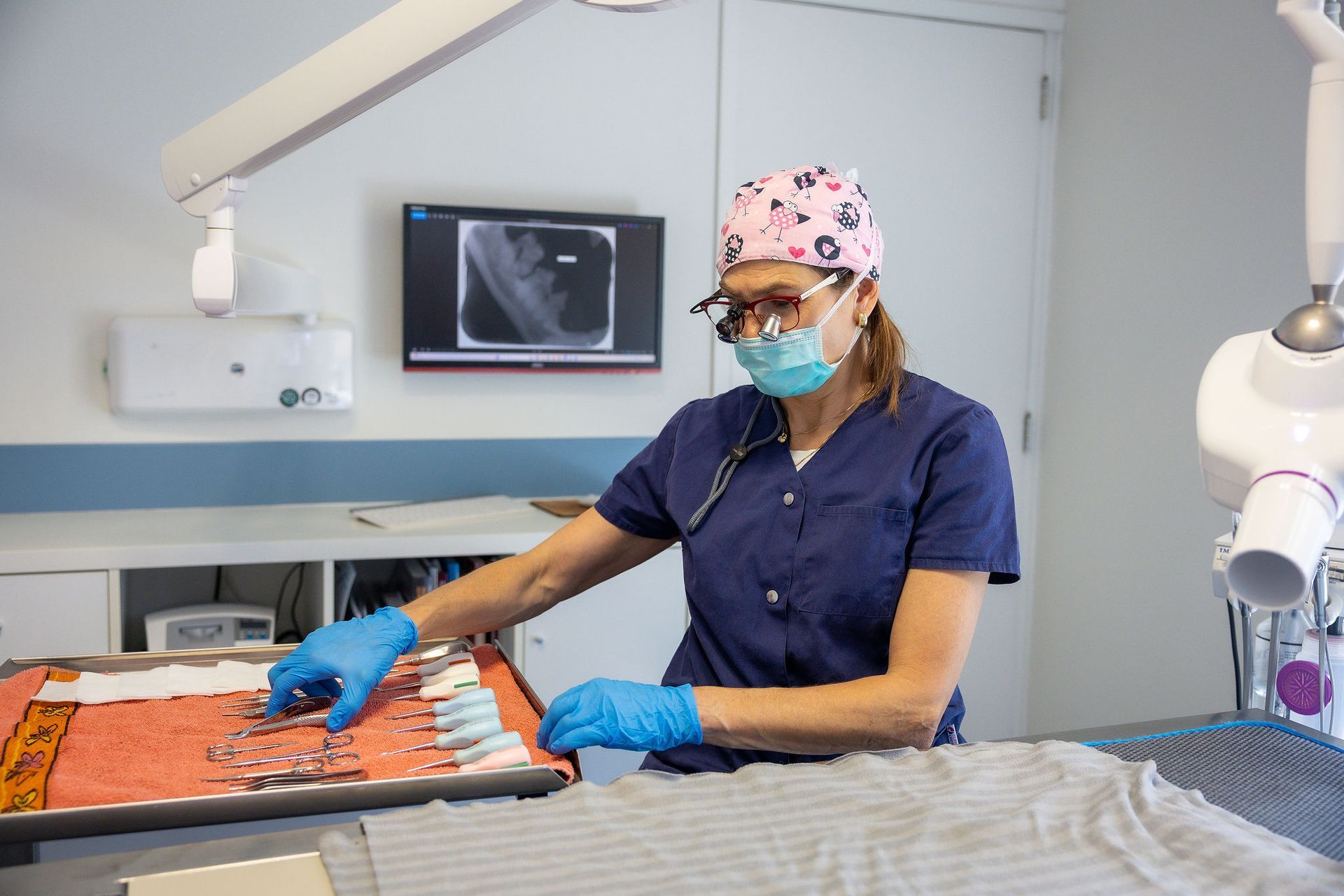
X-rays provide images of the bones, chest or abdomen. They may recommended if your pet is limping, has breathing difficulties or abdominal pain, or if we suspect that they have swallowed a foreign object.
What are ultrasound scans and X-rays?
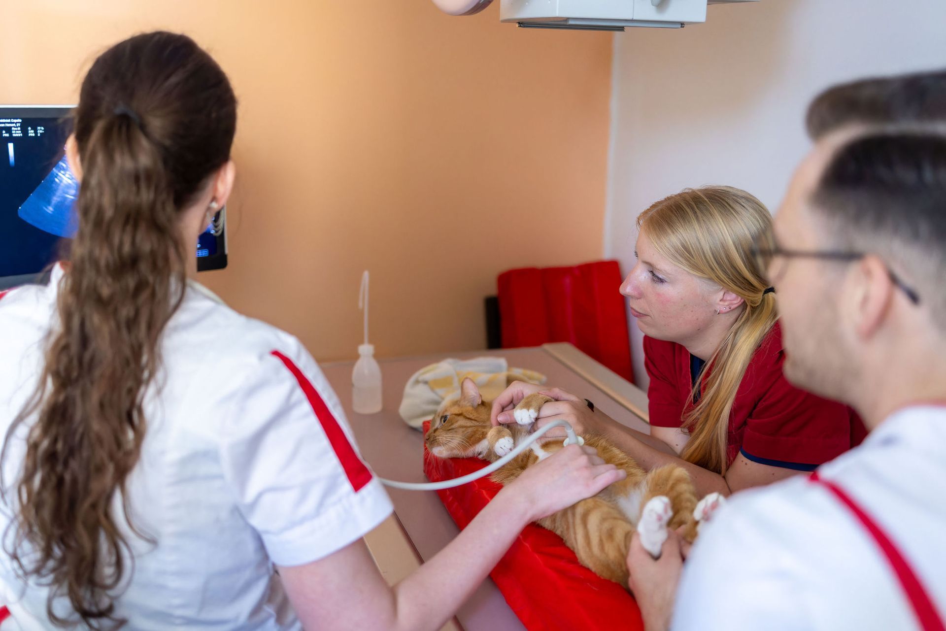
As a veterinary nurse, I work in many different areas of the clinic. From ultrasound and dentistry to the front desk and the operating theatre, I enjoy contributing to the care our team provides for your pet.
Natasja van Hillo
Veterinary Nurse
Sometimes a physical examination does not provide enough information. An ultrasound or X-ray can then help us decide on the most appropriate next step.
Your pet will need to remain very still. In some cases, sedation or a short general anaesthetic may be needed. Sedation helps your pet relax and become sleepy.
Why diagnostic imaging?
Frequently asked questions
What can we examine with an ultrasound scan?
Ultrasound scans are mainly used to examine abdominal organs, such as the liver, kidneys, spleen, gallbladder and bladder. It can also be used to confirm a pregnancy or assess certain masses. For a cardiac ultrasound, we work with a specialist who visits our clinic once a month.
How should I prepare my pet for an ultrasound scan?
For an abdominal ultrasound scan, your pet should not eat for at least eight hours beforehand, but they may drink water. In some cases, we may ask you to prevent your pet from urinating for four hours before the appointment so that their bladder is full. During the scan, your pet lies on a soft cushion.
When is an X-ray recommended?
X-rays can help us identify bone fractures, a foreign object in the stomach or intestines, bladder stones, an enlarged heart or suspected asthma. During pregnancy, X-rays can also help us estimate the expected amount of puppies or kittens.
Are ultrasound scans or X-rays painful for my pet?
Neither procedure is painful. During an abdominal ultrasound scan, your pet needs to lie still on their back. For some X-rays, brief sedation or a short general anaesthetic may be needed so that your pet remains completely still or lie in a position that would otherwise be uncomfortable.
What is radiology and how does it differ from a regular photograph?
Radiology is a general term for imaging tests that provide images of the inside of your pet's body, such as X-rays and ultrasound scans. We choose the test that is most appropriate for your pet's symptoms and discuss it with you beforehand.
Can I take the pregnancy ultrasound images home?
Yes. We can email the pregnancy ultrasound images to you or save them to a USB stick that you bring with you, so you can view them again at home.
What happens if you find something abnormal?
If we find an abnormality, we discuss the possible next steps with you. Sometimes, we can immediately collect a small sample of cells with a fine needle for further testing. In other cases, additional tests or a referral may be needed. We always explain what we know and what remains uncertain.